Research Group Julia Schnabel
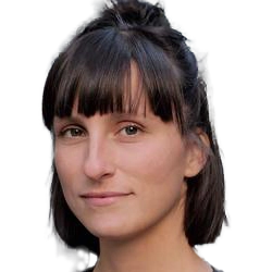
Julia Schnabel
is Professor for Computational Imaging and AI in Medicine at TU Munich.
Her field of research comprises medical image computing and machine learning. Her research focuses on intelligent imaging solutions and computer aided evaluation, including complex motion modelling, image reconstruction, image quality control, image segmentation and classification, applied to multi-modal, quantitative and dynamic imaging.
Team members @MCML
PostDocs
PhD Students
Recent News @MCML
Publications @MCML
2025
[37]
X. Zhang • A. Reithmeir • F. Kögl • R. Braren • J. A. Schnabel • D. M. Lang
MedDIFT: Multi-Scale Diffusion-Based Correspondence in 3D Medical Imaging.
Preprint (Dec. 2025). arXiv
MedDIFT: Multi-Scale Diffusion-Based Correspondence in 3D Medical Imaging.
Preprint (Dec. 2025). arXiv
[36]

C. I. Bercea • J. Li • P. Raffler • E. O. Riedel • L. Schmitzer • A. Kurz • F. Bitzer • P. Roßmüller • J. Canisius • M. L. Beyrle • C. Liu • W. Bai • B. Kainz • J. A. Schnabel • B. Wiestler
NOVA: A Benchmark for Rare Anomaly Localization and Clinical Reasoning in Brain MRI.
NeurIPS 2025 - 39th Conference on Neural Information Processing Systems. San Diego, CA, USA, Nov 30-Dec 07, 2025. To be published. Preprint available. URL
NOVA: A Benchmark for Rare Anomaly Localization and Clinical Reasoning in Brain MRI.
NeurIPS 2025 - 39th Conference on Neural Information Processing Systems. San Diego, CA, USA, Nov 30-Dec 07, 2025. To be published. Preprint available. URL
[35]

A. Reithmeir • V. Spieker • V. Sideri-Lampretsa • D. Rückert • J. A. Schnabel • V. A. Zimmer
From Model Based to Learned Regularization in Medical Image Registration: A Comprehensive Review.
Medical Image Analysis.103854. Nov. 2025. In press. DOI
From Model Based to Learned Regularization in Medical Image Registration: A Comprehensive Review.
Medical Image Analysis.103854. Nov. 2025. In press. DOI
[34]
J. Kiechle • S. M. Fischer • D. M. Lang • C. I. Bercea • M. J. Nyflot • L. Felsner • J. A. Schnabel • J. C. Peeken
TomoGraphView: 3D Medical Image Classification with Omnidirectional Slice Representations and Graph Neural Networks.
Preprint (Nov. 2025). arXiv GitHub
TomoGraphView: 3D Medical Image Classification with Omnidirectional Slice Representations and Graph Neural Networks.
Preprint (Nov. 2025). arXiv GitHub
[33]
H. Y. Kim • J. Li • A. B. Solana • C. M. Pirkl • B. Wiestler • J. A. Schnabel • C. I. Bercea
Learning to reason about rare diseases through retrieval-augmented agents.
Preprint (Nov. 2025). arXiv
Learning to reason about rare diseases through retrieval-augmented agents.
Preprint (Nov. 2025). arXiv
[32]
J. Mayr • A. Reithmeir • M. Di Folco • J. A. Schnabel
Covariance Descriptors Meet General Vision Encoders: Riemannian Deep Learning for Medical Image Classification.
Preprint (Nov. 2025). arXiv
Covariance Descriptors Meet General Vision Encoders: Riemannian Deep Learning for Medical Image Classification.
Preprint (Nov. 2025). arXiv
[31]
L. Shilova • D. Sens • A. Aliyeva • S. Xu • E. Salin • J. Schiefelbein • B. Asani • O. V. Amarie • E. Schneltzer • A. V. Segrè • J. A. Schnabel • N. Cai • B. M. Eskofier • F. P. Casale
REECAP: Contrastive learning of retinal aging reveals genetic loci linking morphology to eye disease.
Preprint (Nov. 2025). DOI
REECAP: Contrastive learning of retinal aging reveals genetic loci linking morphology to eye disease.
Preprint (Nov. 2025). DOI
[30]
S. M. Fischer • J. Kiechle • L. Daza • L. Felsner • R. Osuala • D. M. Lang • K. Lekadir • J. C. Peeken • J. A. Schnabel
Progressive Growing of Patch Size: Curriculum Learning for Accelerated and Improved Medical Image Segmentation.
Preprint (Oct. 2025). arXiv
Progressive Growing of Patch Size: Curriculum Learning for Accelerated and Improved Medical Image Segmentation.
Preprint (Oct. 2025). arXiv
[29]

D. M. Lang • R. Osuala • V. Spieker • K. Lekadir • R. Braren • J. A. Schnabel
Temporal Neural Cellular Automata: Application to modeling of contrast enhancement in breast MRI.
MICCAI 2025 - 28th International Conference on Medical Image Computing and Computer Assisted Intervention. Daejeon, Republic of Korea, Sep 23-27, 2025. DOI GitHub
Temporal Neural Cellular Automata: Application to modeling of contrast enhancement in breast MRI.
MICCAI 2025 - 28th International Conference on Medical Image Computing and Computer Assisted Intervention. Daejeon, Republic of Korea, Sep 23-27, 2025. DOI GitHub
[28]

C. K. Wong • A. N. Christensen • C. I. Bercea • J. A. Schnabel • M. G. Tolsgaard • A. Feragen
Influence of Classification Task and Distribution Shift Type on OOD Detection in Fetal Ultrasound.
MICCAI 2025 - 28th International Conference on Medical Image Computing and Computer Assisted Intervention. Daejeon, Republic of Korea, Sep 23-27, 2025. DOI GitHub
Influence of Classification Task and Distribution Shift Type on OOD Detection in Fetal Ultrasound.
MICCAI 2025 - 28th International Conference on Medical Image Computing and Computer Assisted Intervention. Daejeon, Republic of Korea, Sep 23-27, 2025. DOI GitHub
[27]
S. J. Roughley • J. P. Müller • S. Gao • Z. Gao • M. Ligero • R. Blums • M. Crispin-Ortuzar • J. A. Schnabel • B. Kainz • C. I. Bercea • I. P. Machado
GroundingDINO for Open-Set Lesion Detection in Medical Imaging.
MSB EMERGE @MICCAI 2025 - 2nd MICCAI Student Board Emerge Workshop at the 28th International Conference on Medical Image Computing and Computer Assisted Intervention. Daejeon, Republic of Korea, Sep 23-27, 2025. To be published. Preprint available. URL
[26]
N. Niessen • C. M. Pirkl • A. B. Solana • H. Eichhorn • V. Spieker • W. Huang • T. Sprenger • M. I. Menzel • J. A. Schnabel
INR meets Multi-Contrast MRI Reconstruction.
RIME @MICCAI 2025 - 1st Workshop on Reconstruction and Imaging Motion Estimation at the 28th International Conference on Medical Image Computing and Computer Assisted Intervention. Daejeon, Republic of Korea, Sep 23-27, 2025. DOI
INR meets Multi-Contrast MRI Reconstruction.
RIME @MICCAI 2025 - 1st Workshop on Reconstruction and Imaging Motion Estimation at the 28th International Conference on Medical Image Computing and Computer Assisted Intervention. Daejeon, Republic of Korea, Sep 23-27, 2025. DOI
[25]
C. Liu • Y. Chen • H. Shi • J. Lu • B. Jian • J. Pan • L. Cai • J. Wang • Y. Zhang • J. Li • C. I. Bercea • C. Ouyang • C. Chen • Z. Xiong • B. Wiestler • C. Wachinger • D. Rückert • W. Bai • R. Arcucci
Does DINOv3 Set a New Medical Vision Standard?
Preprint (Sep. 2025). arXiv
Does DINOv3 Set a New Medical Vision Standard?
Preprint (Sep. 2025). arXiv
[24]

H. Eichhorn • V. Spieker • K. Hammernik • E. Saks • L. Felsner • K. Weiss • C. Preibisch • J. A. Schnabel
Motion-robust T2* quantification from low-resolution gradient echo brain MRI with physics-informed deep learning.
Magnetic Resonance in Medicine 95.1. Aug. 2025. DOI
Motion-robust T2* quantification from low-resolution gradient echo brain MRI with physics-informed deep learning.
Magnetic Resonance in Medicine 95.1. Aug. 2025. DOI
[23]
S. Ambekar • D. M. Lang • J. A. Schnabel
Hierarchical Adaptive networks with Task vectors for Test-Time Adaptation.
Preprint (Aug. 2025). arXiv
Hierarchical Adaptive networks with Task vectors for Test-Time Adaptation.
Preprint (Aug. 2025). arXiv
[22]
J. Li • C. Liu • W. Bai • M. Liu • R. Arcucci • C. I. Bercea • J. A. Schnabel
Knowledge to Sight: Reasoning over Visual Attributes via Knowledge Decomposition for Abnormality Grounding.
Preprint (Aug. 2025). arXiv GitHub
Knowledge to Sight: Reasoning over Visual Attributes via Knowledge Decomposition for Abnormality Grounding.
Preprint (Aug. 2025). arXiv GitHub
[21]
J. Li • C. Liu • W. Bai • R. Arcucci • C. I. Bercea • J. A. Schnabel
Enhancing Abnormality Grounding for Vision Language Models with Knowledge Descriptions.
Preprint (Mar. 2025). arXiv GitHub
Enhancing Abnormality Grounding for Vision Language Models with Knowledge Descriptions.
Preprint (Mar. 2025). arXiv GitHub
[20]

C. I. Bercea • B. Wiestler • D. Rückert • J. A. Schnabel
Evaluating normative representation learning in generative AI for robust anomaly detection in brain imaging.
Nature Communications 16.1624. Feb. 2025. DOI GitHub
Evaluating normative representation learning in generative AI for robust anomaly detection in brain imaging.
Nature Communications 16.1624. Feb. 2025. DOI GitHub
[19]

J. Li • T. Su • B. Zhao • F. Lv • Q. Wang • N. Navab • Y. Hu • Z. Jiang
Ultrasound Report Generation With Cross-Modality Feature Alignment via Unsupervised Guidance.
IEEE Transactions on Medical Imaging 44.1. Jan. 2025. DOI
Ultrasound Report Generation With Cross-Modality Feature Alignment via Unsupervised Guidance.
IEEE Transactions on Medical Imaging 44.1. Jan. 2025. DOI
[18]
R. Dorent • R. Khajavi • T. Idris • E. Ziegler • B. Somarouthu • H. Jacene • A. LaCasce • J. Deissler • J. Ehrhardt • S. Engelson • S. M. Fischer • Y. Gu • H. Handels • S. Kasai • S. Kondo • K. Maier-Hein • J. A. Schnabel • G. Wang • L. Wang • T. Wald • G.-Z. Yang • H. Zhang • M. Zhang • S. Pieper • G. Harris • R. Kikinis • T. Kapur
LNQ 2023 challenge: Benchmark of weakly-supervised techniques for mediastinal lymph node quantification.
Machine Learning for Biomedical Imaging 3.Special Issue. Jan. 2025. DOI GitHub
LNQ 2023 challenge: Benchmark of weakly-supervised techniques for mediastinal lymph node quantification.
Machine Learning for Biomedical Imaging 3.Special Issue. Jan. 2025. DOI GitHub
2024
[17]
D. Daum • R. Osuala • A. Riess • G. Kaissis • J. A. Schnabel • M. Di Folco
On Differentially Private 3D Medical Image Synthesis with Controllable Latent Diffusion Models.
DGM4 @MICCAI 2024 - 4th International Workshop on Deep Generative Models at the 27th International Conference on Medical Image Computing and Computer Assisted Intervention. Marrakesh, Morocco, Oct 06-10, 2024. DOI GitHub
[16]
A. Riess • A. Ziller • S. Kolek • D. Rückert • J. A. Schnabel • G. Kaissis
Complex-Valued Federated Learning with Differential Privacy and MRI Applications.
DeCaF @MICCAI 2024 - 5th Workshop on Distributed, Collaborative and Federated Learning at the 27th International Conference on Medical Image Computing and Computer Assisted Intervention. Marrakesh, Morocco, Oct 06-10, 2024. DOI
[15]
R. Osuala • D. M. Lang • A. Riess • G. Kaissis • Z. Szafranowska • G. Skorupko • O. Diaz • J. A. Schnabel • K. Lekadir
Enhancing the Utility of Privacy-Preserving Cancer Classification Using Synthetic Data.
Deep-Breath @MICCAI 2024 - 1st Deep Breast Workshop on AI and Imaging for Diagnostic and Treatment Challenges in Breast Care at the 27th International Conference on Medical Image Computing and Computer Assisted Intervention. Marrakesh, Morocco, Oct 06-10, 2024. DOI GitHub
[14]
J. Kiechle • D. M. Lang • S. M. Fischer • L. Felsner • J. C. Peeken • J. A. Schnabel
Graph Neural Networks: A Suitable Alternative to MLPs in Latent 3D Medical Image Classification?
GRAIL @MICCAI 2024 - 6th Workshop on GRaphs in biomedicAl Image anaLysis at the 27th International Conference on Medical Image Computing and Computer Assisted Intervention. Marrakesh, Morocco, Oct 06-10, 2024. DOI URL
[13]

S. M. Fischer • L. Felsner • R. Osuala • J. Kiechle • D. M. Lang • J. C. Peeken • J. A. Schnabel
Progressive Growing of Patch Size: Resource-Efficient Curriculum Learning for Dense Prediction Tasks.
MICCAI 2024 - 27th International Conference on Medical Image Computing and Computer Assisted Intervention. Marrakesh, Morocco, Oct 06-10, 2024. DOI GitHub
Progressive Growing of Patch Size: Resource-Efficient Curriculum Learning for Dense Prediction Tasks.
MICCAI 2024 - 27th International Conference on Medical Image Computing and Computer Assisted Intervention. Marrakesh, Morocco, Oct 06-10, 2024. DOI GitHub
[12]

A. Reithmeir • L. Felsner • R. Braren • J. A. Schnabel • V. A. Zimmer
Data-Driven Tissue- and Subject-Specific Elastic Regularization for Medical Image Registration.
MICCAI 2024 - 27th International Conference on Medical Image Computing and Computer Assisted Intervention. Marrakesh, Morocco, Oct 06-10, 2024. DOI GitHub
Data-Driven Tissue- and Subject-Specific Elastic Regularization for Medical Image Registration.
MICCAI 2024 - 27th International Conference on Medical Image Computing and Computer Assisted Intervention. Marrakesh, Morocco, Oct 06-10, 2024. DOI GitHub
[11]

T. Su • J. Li • X. Zhang • H. Jin • H. Chen • Q. Wang • F. Lv • B. Zhao • Y. Hu
Design as Desired: Utilizing Visual Question Answering for Multimodal Pre-training.
MICCAI 2024 - 27th International Conference on Medical Image Computing and Computer Assisted Intervention. Marrakesh, Morocco, Oct 06-10, 2024. DOI GitHub
Design as Desired: Utilizing Visual Question Answering for Multimodal Pre-training.
MICCAI 2024 - 27th International Conference on Medical Image Computing and Computer Assisted Intervention. Marrakesh, Morocco, Oct 06-10, 2024. DOI GitHub
[10]
J. Li • S. H. Kim • P. Müller • L. Felsner • D. Rückert • B. Wiestler • J. A. Schnabel • C. I. Bercea
Language Models Meet Anomaly Detection for Better Interpretability and Generalizability.
MMMI @MICCAI 2024 - 5th International Workshop on Multiscale Multimodal Medical Imaging at the 27th International Conference on Medical Image Computing and Computer Assisted Intervention. Marrakesh, Morocco, Oct 06-10, 2024. DOI GitHub
[9]
D. Grzech • L. Le Folgoc • M. F. Azampour • A. Vlontzos • B. Glocker • N. Navab • J. A. Schnabel • B. Kainz
Unsupervised Similarity Learning for Image Registration with Energy-Based Models.
WBIR @MICCAI 2024 - 11th International Workshop on Biomedical Image Registration at the 27th International Conference on Medical Image Computing and Computer Assisted Intervention. Marrakesh, Morocco, Oct 06-10, 2024. DOI
[8]
F. Kögl • A. Reithmeir • V. Sideri-Lampretsa • I. Machado • R. Braren • D. Rückert • J. A. Schnabel • V. A. Zimmer
General Vision Encoder Features as Guidance in Medical Image Registration.
WBIR @MICCAI 2024 - 11th International Workshop on Biomedical Image Registration at the 27th International Conference on Medical Image Computing and Computer Assisted Intervention. Marrakesh, Morocco, Oct 06-10, 2024. DOI URL
[7]

A. C. Erdur • D. Rusche • D. Scholz • J. Kiechle • S. Fischer • Ó. Llorián-Salvador • J. A. Buchner • M. Q. Nguyen • L. Etzel • J. Weidner • M.-C. Metz • B. Wiestler • J. A. Schnabel • D. Rückert • S. E. Combs • J. C. Peeken
Deep learning for autosegmentation for radiotherapy treatment planning: State-of-the-art and novel perspectives.
Strahlentherapie und Onkologie 201. Aug. 2024. DOI GitHub
Deep learning for autosegmentation for radiotherapy treatment planning: State-of-the-art and novel perspectives.
Strahlentherapie und Onkologie 201. Aug. 2024. DOI GitHub
[6]
S. M. Fischer • J. Kiechle • D. M. Lang • J. C. Peeken • J. A. Schnabel
Mask the Unknown: Assessing Different Strategies to Handle Weak Annotations in the MICCAI2023 Mediastinal Lymph Node Quantification Challenge.
Machine Learning for Biomedical Imaging 2. Jun. 2024. DOI GitHub
Mask the Unknown: Assessing Different Strategies to Handle Weak Annotations in the MICCAI2023 Mediastinal Lymph Node Quantification Challenge.
Machine Learning for Biomedical Imaging 2. Jun. 2024. DOI GitHub
[5]
J. Kiechle • S. M. Fischer • D. M. Lang • M. Folco • S. C. Foreman • V. K. N. Rösner • A.-K. Lohse • C. Mogler • C. Knebel • M. R. Makowski • K. Woertler • S. E. Combs • A. S. Gersing • J. C. Peeken • J. A. Schnabel
Unifying local and global shape descriptors to grade soft-tissue sarcomas using graph convolutional networks.
ISBI 2024 - IEEE 21st International Symposium on Biomedical Imaging. Athens, Greece, May 27-30, 2024. DOI
Unifying local and global shape descriptors to grade soft-tissue sarcomas using graph convolutional networks.
ISBI 2024 - IEEE 21st International Symposium on Biomedical Imaging. Athens, Greece, May 27-30, 2024. DOI
[4]
J. Kiechle • S. C. Foreman • S. M. Fischer • D. Rusche • V. Rösner • A.-K. Lohse • C. Mogler • S. E. Combs • M. R. Makowski • K. Woertler • D. M. Lang • J. A. Schnabel • A. S. Gersing • J. C. Peeken
Investigating the role of morphology in deep learning-based liposarcoma grading.
ESTRO 2024 - Annual Meeting of the European Society for Radiotherapy and Oncology. Glasgow, UK, May 03-07, 2024. URL
Investigating the role of morphology in deep learning-based liposarcoma grading.
ESTRO 2024 - Annual Meeting of the European Society for Radiotherapy and Oncology. Glasgow, UK, May 03-07, 2024. URL
[3]
A. Reithmeir • J. A. Schnabel • V. A. Zimmer
Learning physics-inspired regularization for medical image registration with hypernetworks.
SPIE 2024 - SPIE Medical Imaging: Image Processing. San Diego, CA, USA, Feb 18-22, 2024. DOI GitHub
Learning physics-inspired regularization for medical image registration with hypernetworks.
SPIE 2024 - SPIE Medical Imaging: Image Processing. San Diego, CA, USA, Feb 18-22, 2024. DOI GitHub
2023
[2]
V. A. Zimmer • K. Hammernik • V. Sideri-Lampretsa • W. Huang • A. Reithmeir • D. Rückert • J. A. Schnabel
Towards Generalised Neural Implicit Representations for Image Registration.
DGM4 @MICCAI 2023 - 3rd International Workshop on Deep Generative Models at the 26th International Conference on Medical Image Computing and Computer Assisted Intervention. Vancouver, Canada, Oct 08-12, 2023. DOI
Towards Generalised Neural Implicit Representations for Image Registration.
DGM4 @MICCAI 2023 - 3rd International Workshop on Deep Generative Models at the 26th International Conference on Medical Image Computing and Computer Assisted Intervention. Vancouver, Canada, Oct 08-12, 2023. DOI
[1]

M. J. Menten • J. C. Paetzold • V. A. Zimmer • S. Shit • I. Ezhov • R. Holland • M. Probst • J. A. Schnabel • D. Rückert
A Skeletonization Algorithm for Gradient-Based Optimization.
ICCV 2023 - IEEE/CVF International Conference on Computer Vision. Paris, France, Oct 02-06, 2023. DOI
A Skeletonization Algorithm for Gradient-Based Optimization.
ICCV 2023 - IEEE/CVF International Conference on Computer Vision. Paris, France, Oct 02-06, 2023. DOI
©all images: LMU | TUM


